<<< Previous page | | Next page >>>
Visualization and analysis of neural activity
Caenorhabditis elegans is an excellent model organism for studying how nervous system produces specific behaviors. How can we observe activity of neurons in the worm?
Although C. elegans has a transparent body, the activity of its neurons cannot be observed as it is. In addition, its small neuron size makes it difficult to measure electrical activity of neurons through injecting electrodes to cell body. However, development of a variety of "calcium indicators", such as GCaMP and cameleon, enables the optical observation of neuronal activities (Fig. 1).
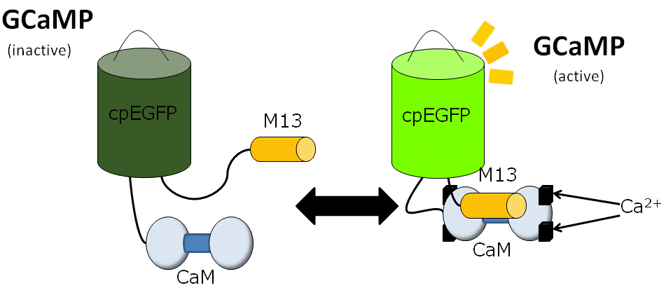
Figure 1: Mechanism of GCaMP. GCaMP is composed of improved GFP, Calmodulin (CaM, calcium-binding protein) and M13 peptide. GCaMP shows fluorescence only when calcium binds to its CaM domain.
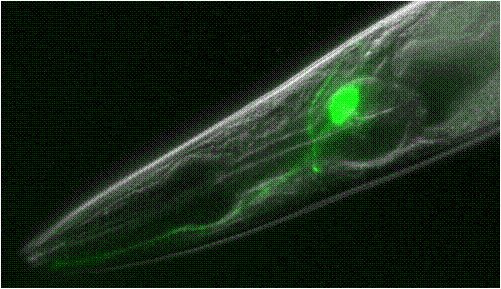
Figure 2: A worm that expresses GCaMP in its ASER sensory neuron.
As a neuron is excited and Ca2+ concentration is increased, fluorescence intensity of the indicator increases and neuronal activity is visualized (Fig. 2). By this method, the plasticity of the nervous system during salt concentration learning was revealed for the first time. Together with genetic study, it is strongly suggested that insulin-like pathway regulates the plasticity (1).
Furthermore, a method to activate specific neurons artificially has been recently developed and called optogenetics. Neurons expressing Channelrhodopsin protein are activated when ultraviolet light is applied. New optogenetic tools are intensively developed and the field is attracting much attentions.
- Shigekazu Oda, Masahiro Tomioka and Yuichi Iino, Neuronal plasticity regulated by the insulin-like signaling pathway underlies salt chemotaxis learning in Caenorhabditis elegans. J Neurophysiol 27 April 2011.
- Satoh et al, Regulation of Experience-Dependent Bidirectional Chemotaxis by a Neural Circuit Switch in Caenorhabditis elegans. J Neurosci. 2014 Nov 19;34(47):15631-7.